Theme 1 : Biosensors
Pierre Vincent; Elvire Guiot

Genetically-encoded biosensors allow real-time imaging of specific intracellular events in living cells. Epac-based sensors (Ponsioen; DiPilato; Nikolaev) report changes in cAMP concentration and AKAR-type biosensors (Zhang) are reporting the PKA/phosphatase equilibrium. These biosensors are expressed in brain slice preparations using viral vectors and imaged in real-time with wide-field (see movie) or two-photon microscopy.

This extremely powerful approach allows us to directly monitor the cAMP/PKA cascade in mature and morphologically intact neurons, and study the integration mechanisms involved in the response to neuromodulators.

Collaborations:
Jin Zhang, Johns Hopkins School of Medicine, Baltimore
Kees Jalink, The Netherlands Cancer Institute, Amsterdam
Fabienne Merola, Université Paris Sud, Orsay
Ludovic Jullien, Département de Chimie, ENS, Paris
With the support of GDR2588.

Theme 2 : Spatial and temporal integration of the cAMP/PKA signal
Elvire Guiot; Liliana Castro; Danièle Tritsch
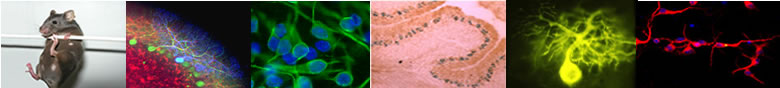
Cortical brain slice expressing the AKAR2.2 biosensor and imaged with two-photon microscopy. Left: Z stack showing individual pyramidal neurons. The color outline shows the region of interest used to perform the ratio calculation indicated by the trace with the corresponding color on the right.
Right: time-lapse recording of the ratio change on the whole stack presented on the left. The pseudo-color image shows one neuron with ratio value coded from blue to red. The ratio change is presented by the thick green trace. The thick grey trace indicates the average response. Drugs are applied in the bath for the duration indicated by the horizontal bars.

Two photon microscopy provides superior spatial resolution allowing us to monitor signal propagation throughout the dendritic tree (figure), while electrophysiology tells us about PKA's effect on membrane channels.
Combining these approaches, we were able to show a spatial compartmentation of the cAMP/PKA signal in neurons, with a "bulk cytosol" domain, where phosphodiesterases maintain a low cAMP level, and a "sub-membrane" domain where cAMP increases faster (Gervasi, 2007) and reaches much higher values (Castro, 2010). We also showed the critical role played by PDE4 in maintaining this spatial compartmentation (Castro, 2010)
We are now analyzing the key factors which control the kinetics and spread of this signal.

Theme 3 : Control of the cAMP/PKA signal
Marina Polito; Liliana Castro

The majority of neurons in the striatum / nucleus accumbens is constituted by medium spiny neurons, whose population is divided in roughly two equal subpopulations: one express dopamine D1-like receptors, positively coupled to the cAMP/PKA cascade and the other subpopulation express D2-like negatively coupled to the cAMP/PKA cascade. These two neuronal populations also differ by the brain regions where their axon projects.
Using cAMP and PKA sensitive probes, we are describing the responses to exogenous dopamine on these two populations. We are studying the functional integration of the dopaminergic signal in these two populations.
We are particularly interested in the functional role played by type 10 phosphodiesterase in the striatum. This enzyme is expressed specifically in this brain region, and new blockers have shown very promising antipsychotic effects, while all other molecules used in clinics to treat schizophrenia share a common antagonistic effect on D2 dopamine receptors. We are trying to understand the link between D2 receptors and PDE10 in the striatum.

Collaborations:
Jean-Antoine Girault and Denis Hervé, Institut du Fer à Moulin

Theme 4 : Functional role in vivo
Pierre Vincent

The cellular approach on brain slice preparations will be paralleled by recordings performed in vivo. In collaboration with Mauna-Kea technologies, fibered fluorescence imaging allowed us to record for the first time calcium signals from neurons located in deep brain regions (Vincent, 2006).
This approach has been improved so as to permit ratiometric imaging in vivo. The fiber is inserted in the nucleus accumbens or in the dorsomedial region of the striatum in anesthetized mice and we are recording the effects of various drugs, injected systemically, which affect dopamine signaling in the striatum.
This new approach will give new clues on the cellular mechanisms involved in addiction. These experiments will eventually lead to recordings in the freely-moving animal, which will then allow a direct monitoring of rewarding events in the nucleus accumbens.

Collaborations:
Mauna-Kea Technologies, Paris